
Several objective clinical tests show whether neuropathy may respond to care. The main ones are nerve conduction studies, electromyography, current perception threshold testing, and intraepidermal nerve fiber density assessment. Each one measures something different about the peripheral nerves. Together, they show how nerves carry signals, how badly a nerve is damaged, and whether its structure can still respond. Nerve conduction studies measure the speed and strength of electrical signals moving through a nerve. Slowed conduction is consistent with nerve damage. Electromyography records electrical activity inside the muscles. It helps separate nerve-based dysfunction from other causes of weakness or pain. Current perception threshold testing quantifies sensory nerve function across different fiber types. It gives a more detailed picture than a standard reflex or pinprick exam. Intraepidermal nerve fiber density testing examines small fiber nerve endings directly. It is especially useful when symptoms point to small fiber neuropathy that standard nerve conduction studies miss. As a group, these tests establish a baseline of remaining nerve function. That baseline does not rely on subjective symptom descriptions alone. The results indicate whether enough functional nerve tissue remains to respond to a structured care plan. They can also show when the degree of nerve damage limits the expected response. Test selection follows the suspected type of neuropathy, the symptom pattern, and findings from an initial neurological examination. These tools are diagnostic methods, not treatments. Their value lies in clarifying the physiological state of the nerve before any care decisions are made. Objective data from these tests replaces guesswork when determining candidacy for nerve-focused care.
Why 'Nothing Shows Up' Doesn't Mean Nothing Is Wrong

You've had the exams, and they came back normal. Your reflexes looked fine and your strength looked fine, yet the burning and numbness never stopped. For so many people with neuropathy, the road to a diagnosis is full of frustration and standard exams that settle nothing.
A clean exam gets treated as proof nothing's wrong. Unexplained doesn't mean untreatable — it means nobody has measured the signal with the right tool yet. That difference decides whether a patient gets sent home or gets a real evaluation, which is exactly what our guide to picking a neuropathy provider walks through.
What Standard Neuropathy Exams Actually Measure
Before anyone mentions advanced testing, most patients have already sat through a standard neurological exam. And it tends to look the same from one office to the next.
That exam isn't worthless. It just measures a much narrower slice of nerve function than most people assume.
The Reflex and Sensation Checklist Most Patients Already Know
You probably know the routine: tapped reflexes, a muscle strength check, and a light touch or pinprick on the skin. Sometimes a tuning fork comes out to test vibration at the toes or ankles.
Those checks screen for damage in the large, heavily myelinated nerve fibers. They'll catch serious motor nerve trouble or major sensory loss, but usually only once it's already advanced.
Why These Checks Stop at the Surface
Here's the problem: small nerve fibers rarely show up under a reflex hammer. Objective nerve function testing gives a clear, data-driven read on what the nerves are actually doing, so the picture doesn't rest on descriptions of pain or numbness alone.
So a standard exam stops at the surface. Its tools were built for large fiber damage, not the small fiber signals that burning feet usually come from. That gap is exactly where using test results to guide neuropathy treatment becomes the next step worth understanding.
What Reflex and Strength Checks Can and Can't Show
Reflex and strength checks were built to find large fiber damage. Small fiber damage often needs a closer look, with a careful sensory exam or a test of the nerve fibers in the skin.
What a Small Fiber Neuropathy Study Actually Found
So how often do these findings show up? A 2021 study by Egenolf et al. in Therapeutic Advances in Neurological Disorders looked at 86 patients with suspected small fiber neuropathy. Abnormal nerve fiber density in the skin of the lower leg was found in 70% of them (60 of 86), and the neurological exam flagged small fiber disease in 62% (53 of 86).
So a careful exam and a skin nerve-fiber test both matter, and neither one tells the whole story alone. That's also why when medication alone stops working for nerve pain becomes the more useful question to ask next.
The Problem With Treating Symptoms Without Nerve Data
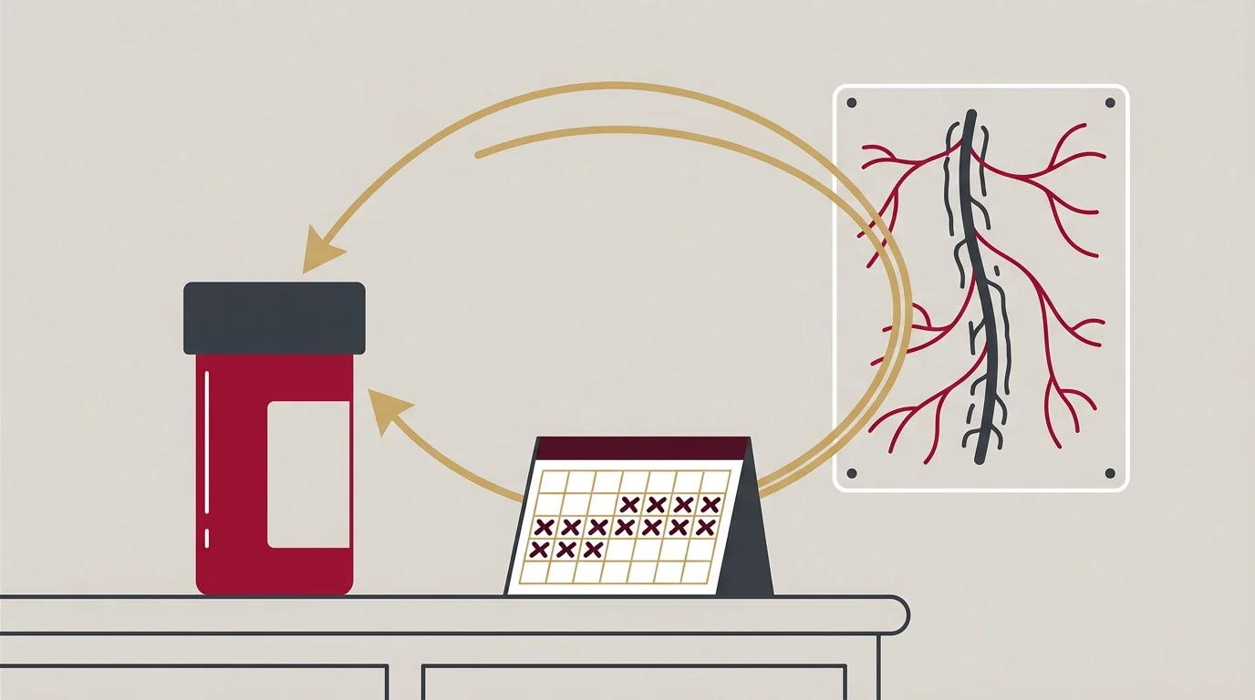
Treating symptoms without nerve data skips the one question that actually matters. The critical question these tests answer is not just what is wrong, but whether enough nerve function remains to allow for a response to care.
A prescription written without that answer is a guess dressed up as a plan.
Open-ended treatment plans built on symptom management alone keep patients in care without ever measuring whether their nerves can respond. A care plan built on precise nerve function data is fundamentally different from one that simply aims to manage symptoms with medication.
Scare tactics and prolonged treatment plans are ethically indefensible. Once nerve function is measured, nervous system focused chiropractic treatment has a baseline to work from instead of a guess to maintain.
How Nerve Conduction and EMG Testing Map Nerve Damage
Nerve conduction studies and needle EMG often get mentioned in the same breath. They measure different things, and mixing them up leads to the wrong test getting ordered.
| Test | What It Measures | Nerve Fiber Type | Who Performs It |
|---|---|---|---|
| Nerve Conduction Study | Speed and strength of electrical signals along larger peripheral nerves | Large, myelinated motor and sensory fibers | Ordered as part of a neurological workup, performed by a trained technician or physician |
| Needle Electromyography | Electrical activity inside muscle tissue at rest and during contraction | Large motor fibers and the muscles they control | A physician, never a chiropractor or other non-physician provider |
| Current Perception Threshold Test | Sensory nerve fiber response across multiple fiber types and stimulus frequencies | Large and small sensory fibers | Ordered as part of a medical nerve workup |
| Intraepidermal Nerve Fiber Density Testing | Density and structure of small nerve fiber endings in the skin | Small, unmyelinated sensory fibers | A provider trained in small fiber neuropathy evaluation, often via skin biopsy analysis |
Where Needle EMG Fits — and Where It Doesn't
Needle EMG places a fine electrode into muscle tissue to read electrical activity at rest and during contraction. The American Association of Neuromuscular & Electrodiagnostic Medicine says chiropractors and other non-physicians don't have the training to perform or interpret needle EMG. So EMG results come from a physician's workup, and the practice reviews them when a patient brings them in.
How Small Fiber and Autonomic Testing Catch What EMG Can't
EMG and nerve conduction studies both focus on larger nerve fibers. Small fiber damage, the kind behind burning or tingling with a normal strength exam, needs a different lens entirely. That's the same fiber-density gap already on the table from the German cohort findings. Testing built for small fiber and autonomic nerve function picks up exactly what EMG was never designed to see.
How Clinical Test Findings Shape a Sequenced Care Plan
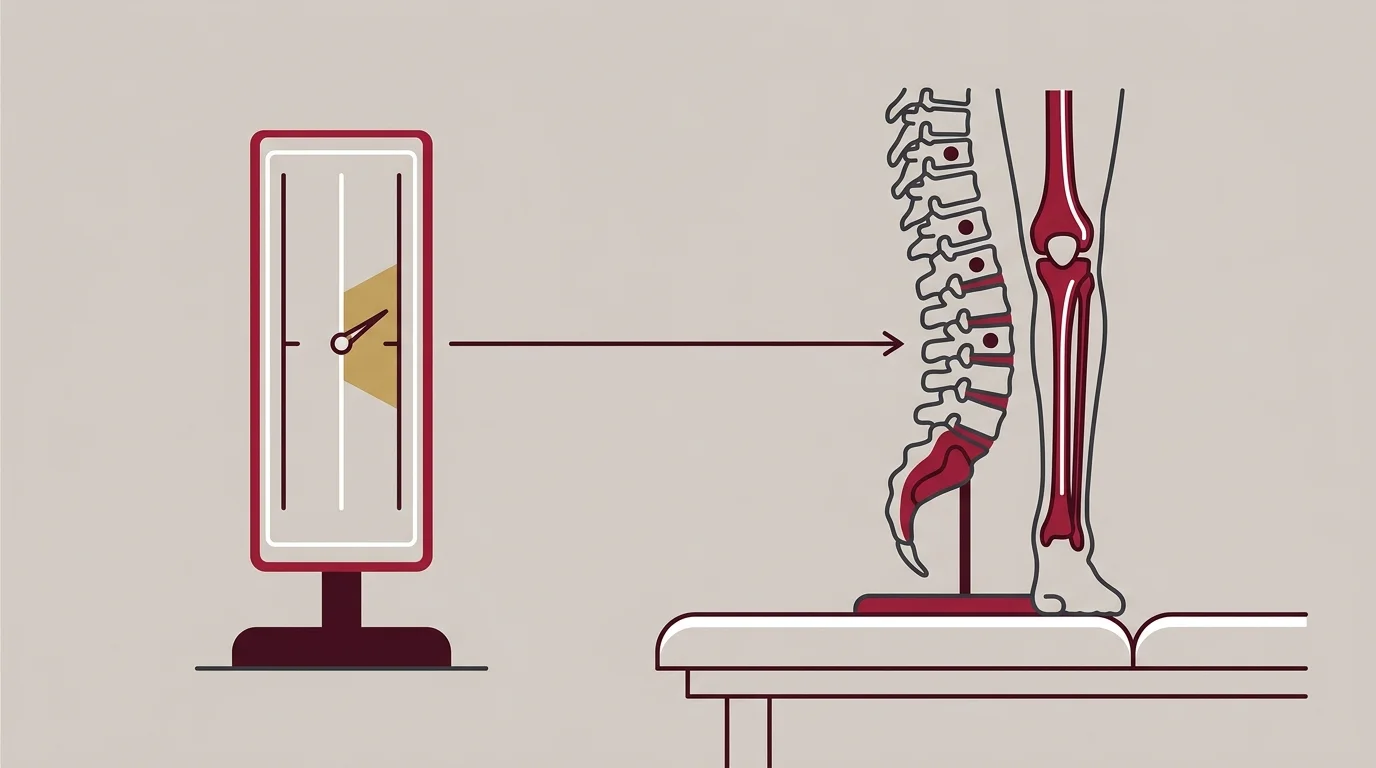
A positive test result is not the finish line. It is the point where results get translated into a sequenced plan, built around what the nerve can still do.
| Step | Test Finding | Resulting Care Decision |
|---|---|---|
| Step 1: Establish Baseline | Testing confirms measurable nerve dysfunction consistent with reported symptoms. | Symptoms are validated as a physiological finding, not dismissed as unexplained. |
| Step 2: Localize the Damage | Results point to small fiber involvement, large fiber involvement, or a radiculopathy pattern. | The suspected source narrows, directing which structures a care plan should target first. |
| Step 3: Gauge Remaining Function | Findings show functional nerve tissue still present, even where damage is significant. | Chiropractic adjustment and nerve-focused care are considered viable, with a defined starting point. |
| Step 4: Identify Limited Response Potential | Findings show nerve damage severe enough to limit expected functional recovery. | Expectations are set honestly, and referral or a different care pathway is discussed. |
| Step 5: Sequence and Reassess | Initial results set a baseline against which later retesting can be compared. | Care plan adjustments are based on measured change, not on how a patient feels that week. |
Reading Nerve Fiber Density and Perception Threshold Results
No protocol is proven for every patient. That is why reading intraepidermal nerve fiber density and current perception threshold results case by case matters more than running the same fixed approach on every presenting complaint.
A 1991 report by Hill and Lawrence in the Journal of the American Podiatric Medical Association described two foot-pain cases where current perception threshold testing supported a suspected nerve root problem.
That kind of finding is exactly why test data, not a standard protocol, decides the next step.
Building a Plan Around What the Nerves Can Still Do
Reading the results is step one. Step two is sequencing a plan around them. That starts with how much working nerve tissue the testing confirmed is still there.
Frequently Asked Questions
Some questions come up more than the rest. So here are the straight answers.
How do these specialized tests differ from the nerve tests my family doctor performed?
A family doctor's exam usually checks reflexes, strength, and basic sensation. These tests measure nerve fiber density, sensory thresholds, and conduction speed directly, which adds detail a quick exam can't give.
Are the clinical tests for neuropathy painful or invasive?
Most of these tests are minimally invasive. Sensory threshold testing involves no needles at all, and nerve fiber density assessment uses a small skin sample rather than surgery.
What happens if testing shows nerve damage is too severe to respond to care?
Severe nerve damage changes what a plan can realistically target. The data still has value here, because it points toward the right next referral instead of continuing a guess.
Can these tests pinpoint the exact cause of neuropathy?
These tests confirm how much nerve function remains and where the damage pattern points. They narrow the picture considerably, though the underlying cause sometimes requires additional medical workup to confirm fully.
Does everyone with neuropathy symptoms need this level of testing first?
Not every case needs the full battery. Symptom pattern and findings from an initial neurological exam determine which tests, if any, are the right next step.
Where This Leaves the Diagnosis
An unexplained symptom is not proof there's nothing wrong. It is proof nobody has measured it with the right instrument yet.
That's the line this whole diagnostic picture rests on. A normal reflex test closes a chart, while a current perception threshold result, a nerve fiber density finding, or a nerve conduction study opens it right back up.
Guesswork treats symptoms. Measurement treats nerves. For patients dismissed by traditional medicine, the nerve function data is the first real answer, and the next step is finding out what that data says about your own case through a consultation with Touch Of Wellness Chiropractic.
Results may vary.
Insurance coverage varies by plan and should be verified before your visit.